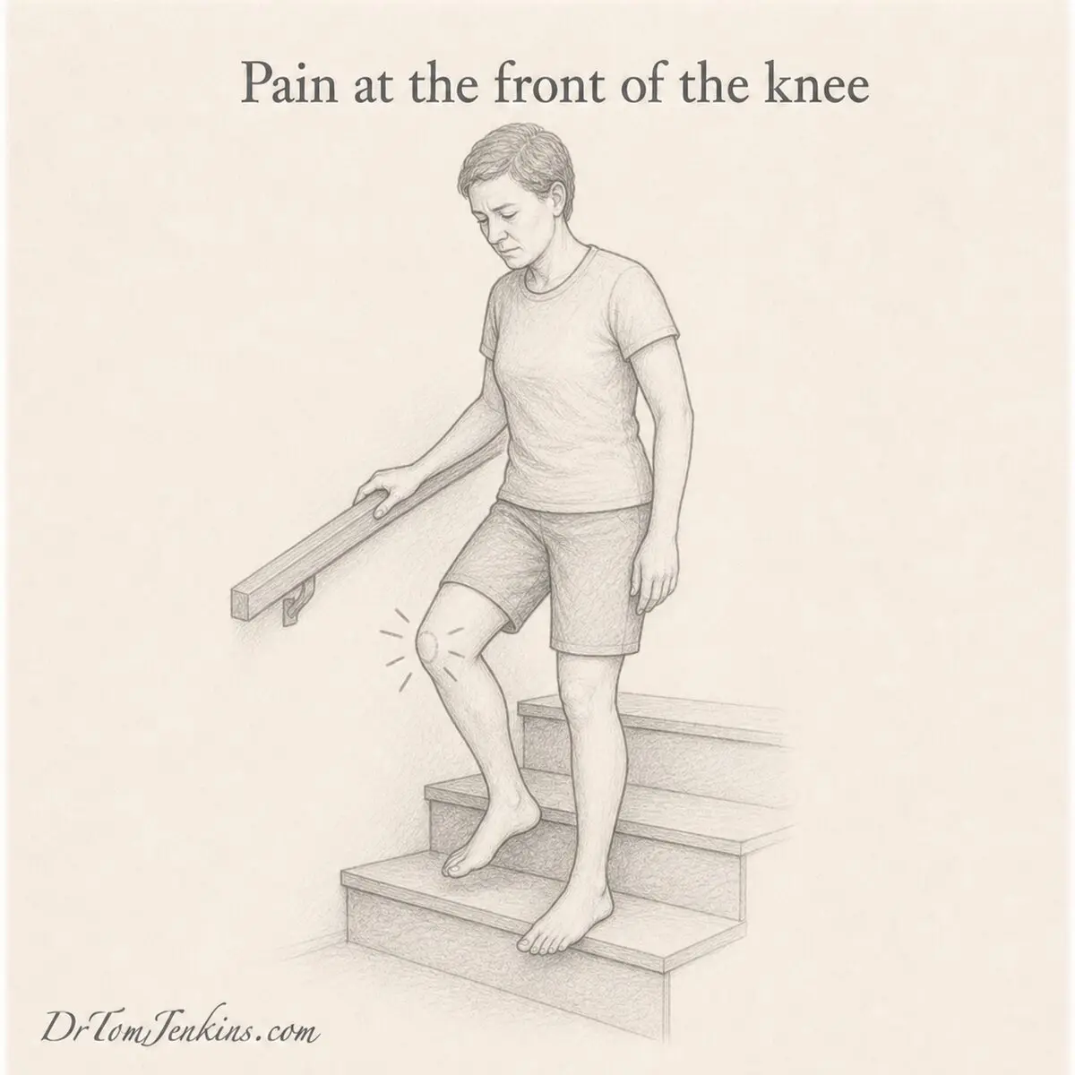
Many people come to my clinics in Gloucestershire describing pain at the front of the knee, or kneecap pain. It flares going up or down stairs, walking downhill, squatting, kneeling or running, and it aches after sitting for a long time with the knee bent, the classic discomfort when you stand up at the end of a film. It is one of the most common problems I see, and one of the most misunderstood.
Two things are worth saying at the outset. It is rarely a sign that the knee is damaged. And for most people it can be settled, though it usually takes longer than they expect.
This article covers anterior knee pain as a whole, with the main focus on its commonest cause, patellofemoral pain. It explains what is going on, why the answer is genuinely different for different people, what helps and why, and it finishes with a home exercise programme you can start straight away while you are waiting to be seen.
In a hurry? Jump straight to the home exercise programme ↓
What “anterior knee pain” actually means
Anterior knee pain is an umbrella term, not a diagnosis. It simply means pain at the front of the knee, and several different structures can produce it.
Patellofemoral pain is by far the commonest. This is pain arising from the joint between the kneecap (patella) and the thigh bone (femur) behind it. It is also called anterior knee pain, runner’s knee, or the older term chondromalacia patellae.
Patellar tendinopathy, sometimes called jumper’s knee, affects the tendon running from the bottom of the kneecap to the shin. Quadriceps tendinopathy affects the tendon just above the kneecap. Fat pad irritation involves the soft, sensitive pad of tissue just behind and below the kneecap. Excessive lateral pressure and patellofemoral instability involve the kneecap sitting or running too far towards the outer side, or slipping out of its groove altogether.
In younger people, Osgood-Schlatter disease and Sinding-Larsen-Johansson syndrome cause pain at the growing attachment points. In older people, patellofemoral osteoarthritis becomes more relevant. Occasionally, pain felt at the front of the knee is actually referred from the hip or the back, which is why the hip should always be examined.
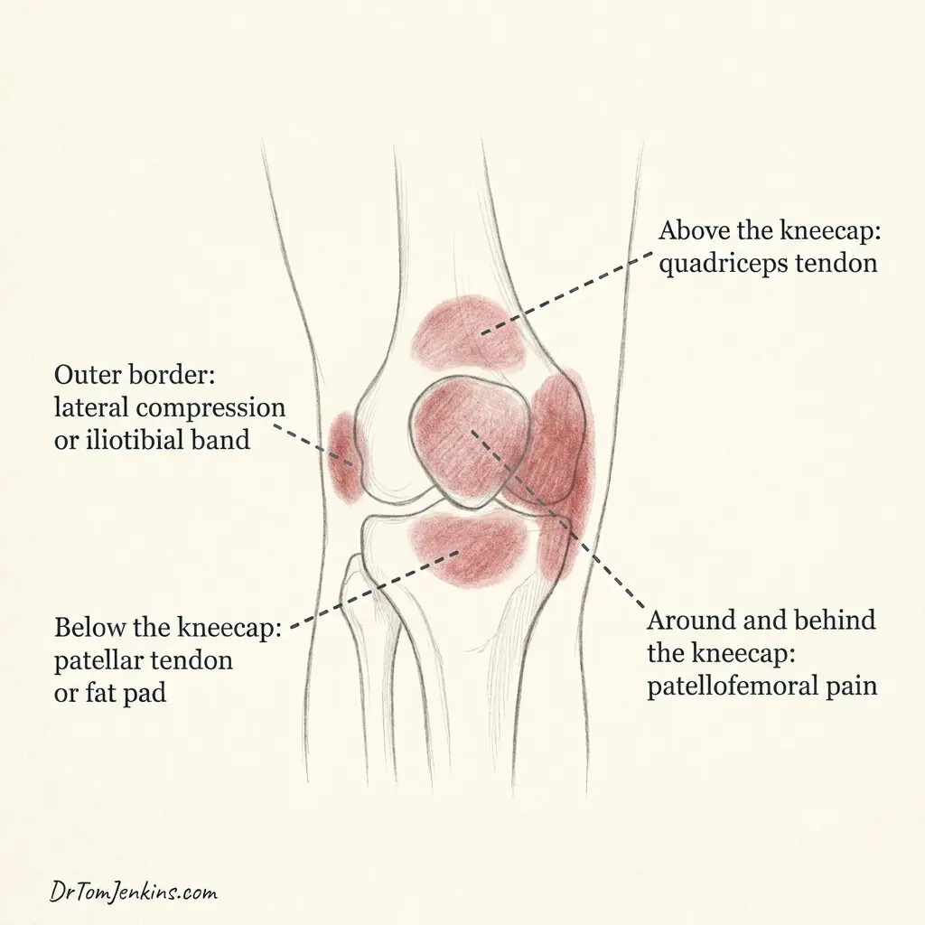
Where it hurts is one of the most useful clues. A diffuse ache that is hard to point to, felt around or behind the kneecap, suggests the patellofemoral joint. Pain pinpointed just below the kneecap suggests the patellar tendon or the fat pad. Pain just above the kneecap suggests the quadriceps tendon. Pain concentrated along the outer border suggests excessive lateral pressure or the iliotibial band. These conditions can and often do coexist, which is part of why careful assessment matters.
The rest of this article concentrates on patellofemoral pain, but most of the general principles, and the exercise programme at the end, apply sensibly across the whole group.
How the knee is built
I often describe the knee as two poles stacked one on top of the other, with your body weight balanced above. Where they meet has to bend, so unlike movement side to side or backwards, which is limited by strong ligaments, the bending movement at the front is controlled almost entirely by muscle.
The large thigh muscles at the front, the quadriceps, join together into a single strong strap that runs down over the kneecap and attaches to your shin. The kneecap sits in a shallow groove at the end of the thigh bone, called the trochlea, and glides up and down within that groove as you bend and straighten.
The kneecap is held on that path by a balance of pull from either side. On the inner side, the vastus medialis draws it inwards. On the outer side, the vastus lateralis, together with the iliotibial band running down the outside of the thigh, draws it outwards.
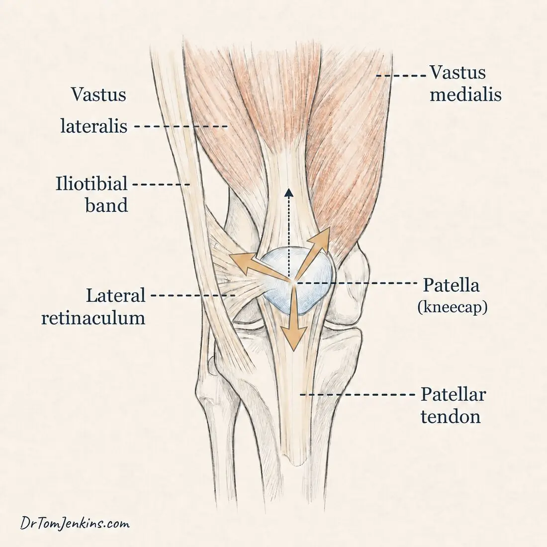
That band is worth knowing about for two reasons. It runs from the pelvis to the outer side of the shin, and the tensor fasciae latae and part of the gluteus maximus attach into it, so it is the route through which the muscles of the hip and buttock act on the knee. This is one of the reasons hip strength matters so much for a problem felt at the front of the knee.
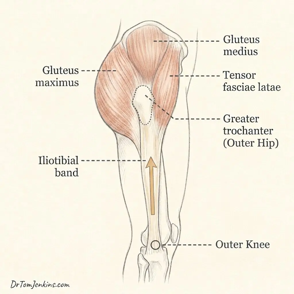
It also connects directly to the kneecap, through a sheet of fibrous tissue on the outer side called the lateral retinaculum. That is the anatomical reason why tightness on the outside of the thigh can influence how the kneecap sits and moves. For that system to work comfortably, the muscles need to be strong enough and coordinated enough to control the kneecap and steady the knee under load: not only the thigh muscles, but the muscles at the side and back of the hip, the glutes, which control how the whole thigh lines up beneath the kneecap when you put weight through a bent knee.
Why kneecap pain hurts
Every tissue has a certain capacity: an amount of load it can take before it complains. Pain at the front of the knee usually appears when the load going through the joint exceeds what it can currently tolerate. The tissue becomes irritated and sensitive.
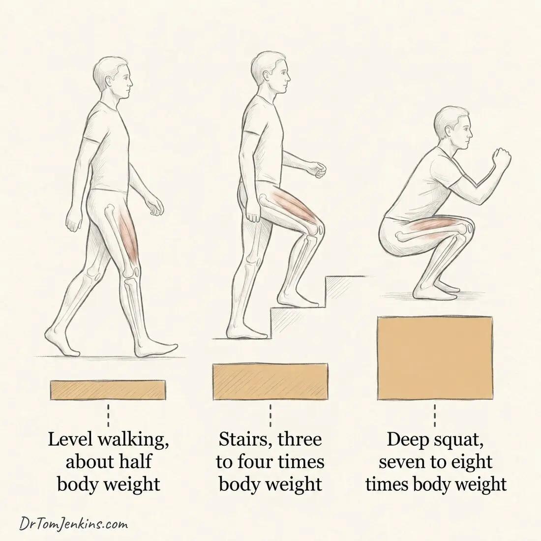
That is why the pain follows load so faithfully. The kneecap joint is under least pressure when the leg is straight, and most pressure when the knee is bent under body weight.
The numbers are striking. Walking on level ground puts roughly half your body weight through the kneecap joint. Going up and down stairs puts three to four times your body weight through it. A deep squat can reach seven or eight times. That is why a knee which feels fine on the flat can become sore within one flight of stairs, and why fairly modest changes in how much of that loading you do can make such a difference.
Why the picture is different for different people
This is the part I feel most strongly about, and it is where honest disagreement still exists between clinicians.
There is an ongoing debate about how much things like the way the kneecap tracks, tightness in the iliotibial band and the structures on the outer side of the kneecap, and the balance and timing of the thigh muscles genuinely matter. Some research finds these features just as often in people with no pain at all, and finds that treatments which relieve pain often do so without measurably changing them. Other well-respected and entirely current clinical sources still place them at the centre of the problem. Both positions are held by thoughtful people looking at the same evidence.
My own view, formed over many years of assessing these knees, is that the importance varies from person to person. In one patient the dominant issue really is tightness and compression on the outer side of the kneecap. In another it is weak hip control allowing the thigh to roll inwards, so the kneecap is loaded awkwardly on every step. In a third the knee is built and controlled perfectly well, and they simply did too much too quickly. The factor that matters enormously in one person is irrelevant in the next.
This is exactly why a thorough clinical assessment matters, and it is probably the single most useful thing in this article. If you follow a generic programme aimed at a problem you do not actually have, you can work hard for months and get nowhere. Not because the treatment was bad, but because it was answering someone else’s question. The exercises later are a safe starting point for almost anyone with pain at the front of the knee. But if you are doing them faithfully and not improving, the answer is usually not to try harder. It is to be assessed properly, so that the plan is aimed at what is actually driving your pain.
What that assessment actually involves. Much of it is simply watching you move, on one leg and under load, because that is when the pattern tends to show itself. Alongside that we look at the strength and control through the thigh and the hip, and at the history that led up to it: what changed, and when.
The most useful part is often what happens when we change something. If altering how you are moving, or how much load is going through the joint, changes your pain there and then, that is a strong clue about where to start. And it is worth saying plainly that a knee rolling inwards, or a pelvis dropping, is not automatically a fault. Plenty of people move that way with no pain whatsoever. These are clues to be tested, not defects to be corrected on sight.
The factors that may be contributing
Any of the following may be relevant to you, and part of the assessment is working out which.
Training and activity load. A jump in running, a new gym routine, a long walk on holiday, a house move. Often the whole story, particularly when the knee is otherwise well built.
Quadriceps capacity and control. How strong the thigh is, and how well it works. One long-standing idea is that if the muscle on the inner side is slow to switch on compared with the one on the outer side, the kneecap is guided less well. This remains part of the mainstream clinical picture, though it is a correlation rather than a proven cause and is one of the more debated points. The practical conclusion is settled enough: the quadriceps is trained as a whole, because attempts to isolate one part of the muscle have not been shown to work better than general thigh strengthening.
Hip and buttock control. Weakness of the muscles that take the leg out to the side and rotate it outwards allows the thigh to drop and turn inwards under load.
Trunk and pelvic control. The knee is the far end of a chain. How well you hold the pelvis and trunk steady on one leg changes what arrives at the kneecap.
Tightness on the outer side. The iliotibial band and the lateral structures around the kneecap can be tight and tender, and in some people this appears to be a genuine driver.
Kneecap position and mobility. Some kneecaps sit or run slightly towards the outer side, or are unusually stiff or unusually mobile.
Ankle movement. If the ankle does not bend forward easily, the knee and hip have to absorb more of the load in a squat or a step down.
Foot posture. Feet that roll inwards markedly can alter how the whole leg lines up.
Previous injury and general conditioning. An old injury, a spell of inactivity or illness, or simply being less conditioned than the activity demands, all lower the load the joint will accept before it complains.
Body weight. Because the force through the joint is a multiple of body weight, losing weight reduces load where it is relevant. It is worth being clear that carrying extra weight has not been shown to cause this problem in the first place, so this is about reducing load rather than assigning blame.
Almost nobody has all of these. The point of the assessment is to find the two or three that matter in your knee, and to leave the rest alone.
The myths worth clearing up
“It is wear and tear, or bone on bone.” Your cartilage may or may not show some wear, and if you have had a scan you may well have been told that it does. Either way, that wear is not what is directly causing your pain. The cartilage surfaces have no nerve endings and cannot themselves feel pain, and scans of completely pain-free knees very often show the same changes as painful ones.
“The clicking and grinding means damage.” That noise, crepitus, is extremely common and is found in most pain-free knees. It is not a sign that harm is being done when you move.
“Rest is the cure.” Complete rest reduces the load, which helps briefly, but it also reduces capacity, so the problem returns as soon as you resume. The aim is to lower load temporarily while building capacity.
Do I need a scan?
Usually not, at least not to explain the pain itself. A scan cannot diagnose patellofemoral pain, and because harmless age-related changes are so common, imaging often shows findings that have nothing to do with your symptoms and can cause needless alarm. Where imaging earns its place is in answering a different question: whether something else is going on as well, or instead, such as patellar tendinopathy, a cartilage or meniscal problem, or the features associated with an unstable kneecap. It is there to check for co-existing conditions when the history or examination raises the question, not to explain the pain.
The simplest way I can put it is this: a scan shows the parts, and the assessment shows how those parts are working together. Two people can have almost identical pictures, one of them struggling to manage a flight of stairs and the other running without a thought. That gap is the whole reason the examination matters, and it is why a report on its own cannot tell you what your rehabilitation should look like.
None of which makes imaging unimportant. When there is a genuine question about the structure of the joint, it answers it well, and it is worth requesting one when that question arises.
Other labels you may be given
Patients are often given a slightly different name by different clinicians, which is understandably confusing. These are best thought of as points along one spectrum rather than separate diseases.
At the common end is patellofemoral pain as described above, in a knee built entirely normally, with a diffuse ache that is hard to pinpoint. Further along is excessive lateral pressure, where the kneecap runs slightly too far towards the outer side so its outer edge is pressed harder than it should be: the pain is concentrated along the outer border, and the kneecap often feels tight and does not glide inwards easily, with no sense of it slipping. At the far end is patellofemoral instability, where the kneecap slips partly or fully out of its groove. Here the story changes: instead of a load-related ache there is a sense of the kneecap shifting sideways or giving way, sometimes needing to be put back, often with swelling afterwards.
What they share matters as much as what separates them. All are aggravated by stairs, hills, squatting, kneeling and prolonged sitting, all are load-sensitive, and they can coexist. And the foundation of treatment is the same: settle the load, then progressively rebuild strength and control in the thigh and hip. The differences are of emphasis. So if you have been given one of these labels rather than another, it does not mean you have a different or more serious disease.
If you have been told you have arthritis behind the kneecap
Wear behind the kneecap becomes commoner with age, and some people reading this will have been told they have patellofemoral osteoarthritis. That is a different thing from the load-related pain described above, although the two often sit together in the same knee.
The message I most want to land is that a diagnosis of arthritis does not fix your symptoms in place. Exercise will not reverse the structural change, and I am not going to pretend otherwise. But how much pain that joint gives you, and how much it stops you doing, are far more movable than the report tends to suggest. Managing load sensibly, building strength through the thigh and hip, and improving general fitness all reduce symptoms in arthritic joints, which is why NICE recommends tailored exercise for everyone with osteoarthritis rather than saving it for the mild cases.
Surgery does have a place, but it is for a minority: people with severe symptoms who have given proper non-operative treatment a fair run and are still struggling. It is not the automatic next step after a scan report.
Will it get better, and how long will it take?
Most people can settle their kneecap pain, but I would rather be straight about the timescale, because unrealistic expectations are one of the main reasons people give up too soon. This is a matter of months rather than weeks, and it is normal to see little change in the first few weeks before things begin to turn.
The follow-up studies give a mixed picture. Roughly four in ten people still have symptoms a year after standard treatment, so this is not a problem that reliably disappears on its own. Set against that, when people were followed up seven years later, around eight in ten of those who had kept going with a home exercise programme rated their outcome as good. That contrast is the important part. The people who do best are those who understand the problem, manage their load sensibly, and keep the strengthening going after the pain has settled.
What helps with kneecap pain, and why
Understanding it and managing load. Ease off, but do not stop, the activities that most aggravate it, and spread loading rather than doing it in spikes. This brings load below the level that irritates the joint so it can settle. Swimming, cycling and cross-training usually let you keep fitness meanwhile.
Strengthening the hip and thigh. This is the best-supported treatment by a wide margin, and it is the main event. A progressive programme working both the thigh and the muscles at the side and back of the hip restores capacity and control. Early on, hip-focused work is often more comfortable and is a good place to start.
Addressing what is specific to you. Stretching or releasing tight lateral structures, attention to foot posture, or adjusting running technique, where the assessment shows these are relevant.
Adjuncts that buy a window. Taping the kneecap, tailored to your particular knee, can reduce pain over the first few weeks. Simple off-the-shelf insoles help some people, particularly those whose feet roll inwards markedly. Both work best alongside a strengthening programme rather than instead of one.
Settling the pain buys you time; building strength buys you the future.
Home exercises to start now
These exercises can be used while you are waiting to see a physiotherapist. They are a safe and sensible starting point for most people with pain at the front of the knee, whichever of the anterior knee conditions is responsible. They do not replace physiotherapy: a physiotherapist examines the whole leg in person, works out which factor is actually driving your pain, corrects your technique, judges when you are ready to progress, and changes the plan when it is not working, which is the part no written programme can do.
Please read this first. These are general exercises for people with a confirmed or reasonably clear diagnosis of patellofemoral pain or a similar load-related anterior knee problem, not a personalised programme. Do not start them if your knee locks or gives way, is significantly swollen, or if the pain followed a distinct injury. If you are unsure of the diagnosis, get assessed first. And if you have worked through the early stages consistently for six to eight weeks and are no better, do not simply push harder: that is precisely the point at which you need a proper assessment.
Start here: two exercises to build into your day
If you take only one thing from this article, take this. I would far rather you did two simple exercises every single day for a year than a sophisticated programme once a fortnight. Consistency beats complexity, comfortably. So begin with these two, and attach each one to something that already happens in your day.
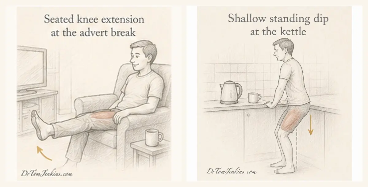
Seated knee extensions, during the advert breaks. Sitting in your usual chair, straighten the knee out in front of you, hold for a couple of seconds, then lower it slowly. Keep going through the first two or three advert breaks of whatever you are watching in the evening. No weight and no equipment. If the last part of straightening is uncomfortable, work in the middle of the range and stop short of fully straight.
Shallow standing dips, while the kettle boils. Standing at the kitchen worktop with both hands resting on it, bend your knees a small way, then straighten up. Shallow and controlled, letting the worktop take some of your weight. Every time you wait for the kettle.
The point of tying these to the advert break and the kettle is that those things happen anyway. They become the reminder, so you are not relying on willpower or on remembering. It is the same reason you brush your teeth every day without deciding to: it is welded to an existing routine. The behavioural research supports this squarely, and it takes a median of around two months of daily repetition before something becomes genuinely automatic. Missing the occasional day does not undo the progress.
The full exercise programme
The fuller programme below will give you a better chance of recovery, and if you have the time it is well worth doing. It works through the thigh, the hip and the buttock in stages, and adds stretches. It is adapted from the patient exercise leaflet shared by the British Patellofemoral Society, developed by senior physiotherapist Celia Wogan and the Bristol Knee Group at University Hospitals Bristol, and cross-checked against NHS physiotherapy leaflets. I am grateful to them for making it freely available.
The rules. Do them little and often, most days. The pain rule: some discomfort during and after exercise is expected and acceptable. Your pain should not rise by more than about a fifth above its usual level for more than an hour afterwards, and it should be no worse the next morning. If it is, reduce the repetitions or the depth, not the frequency. Progression: when you can manage 25 to 30 repetitions comfortably with good technique, move on to the next stage. And do not expect quick change: give it six to twelve weeks before judging it. If the early exercises feel too easy to be doing anything, that is deliberate, and there is more on why further down.
Stage 1: settling the knee
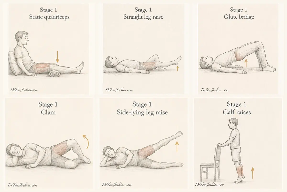
Static quadriceps. Sit with the leg straight and a rolled towel under the knee. Press the back of the knee down into the towel, tightening the thigh. Hold 10 seconds, repeat 10 times. Straight leg raise. Lie on your back, one knee bent, the affected leg straight; tighten the thigh, lift the straight leg about 20cm, lower slowly, keeping the knee locked straight. Glute bridge. Lie on your back, knees bent, feet slightly wider than hips; squeeze your buttocks and lift your hips without arching your back. Clam. On your side, knees bent to 90 degrees, heels together; lift the top knee towards the ceiling without rolling your pelvis backwards. Side-lying leg raise. On your side, bottom leg bent, top leg straight; lift it with the toes pointing forwards. Calf raises. Holding a support, rise onto your toes and lower slowly.
Stage 2: building capacity
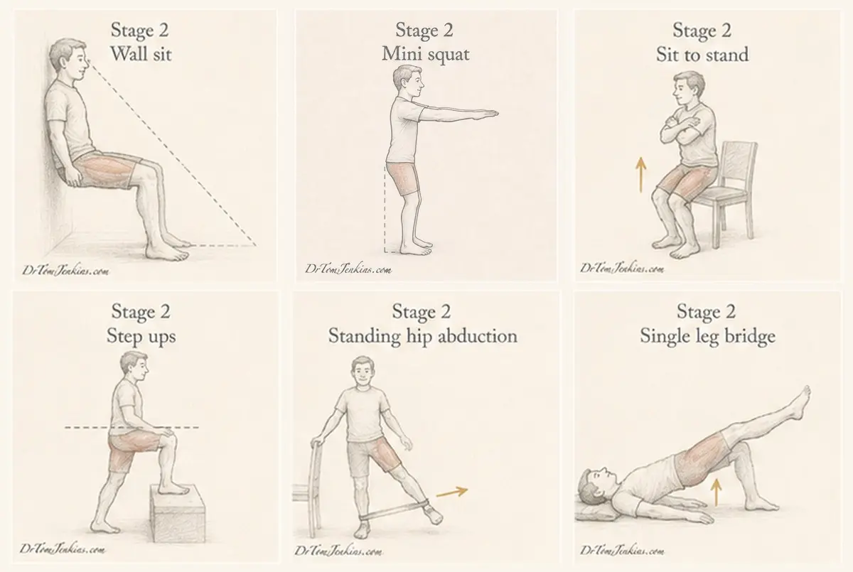
Wall sit. Back against a wall, feet 30cm out and hip width apart, knees bent to about 45 degrees so you can still see your toes; hold 10 seconds initially, building up. Mini squat. A shallow squat, no more than about 30 degrees of knee bend, pushing your bottom back and keeping your knees tracking over your feet. Sit to stand. From a firm chair, stand and sit slowly without using your hands, keeping the knees from rolling inwards. Step ups. Start with a low step, step up with the affected leg, keeping the pelvis level and the knee tracking forwards. Standing hip abduction. Standing tall holding a support, take the leg out to the side keeping the pelvis level; progress by adding a resistance band. Single leg bridge. As the glute bridge, but with the unaffected leg held straight off the floor.
Stage 3: loading and returning to activity
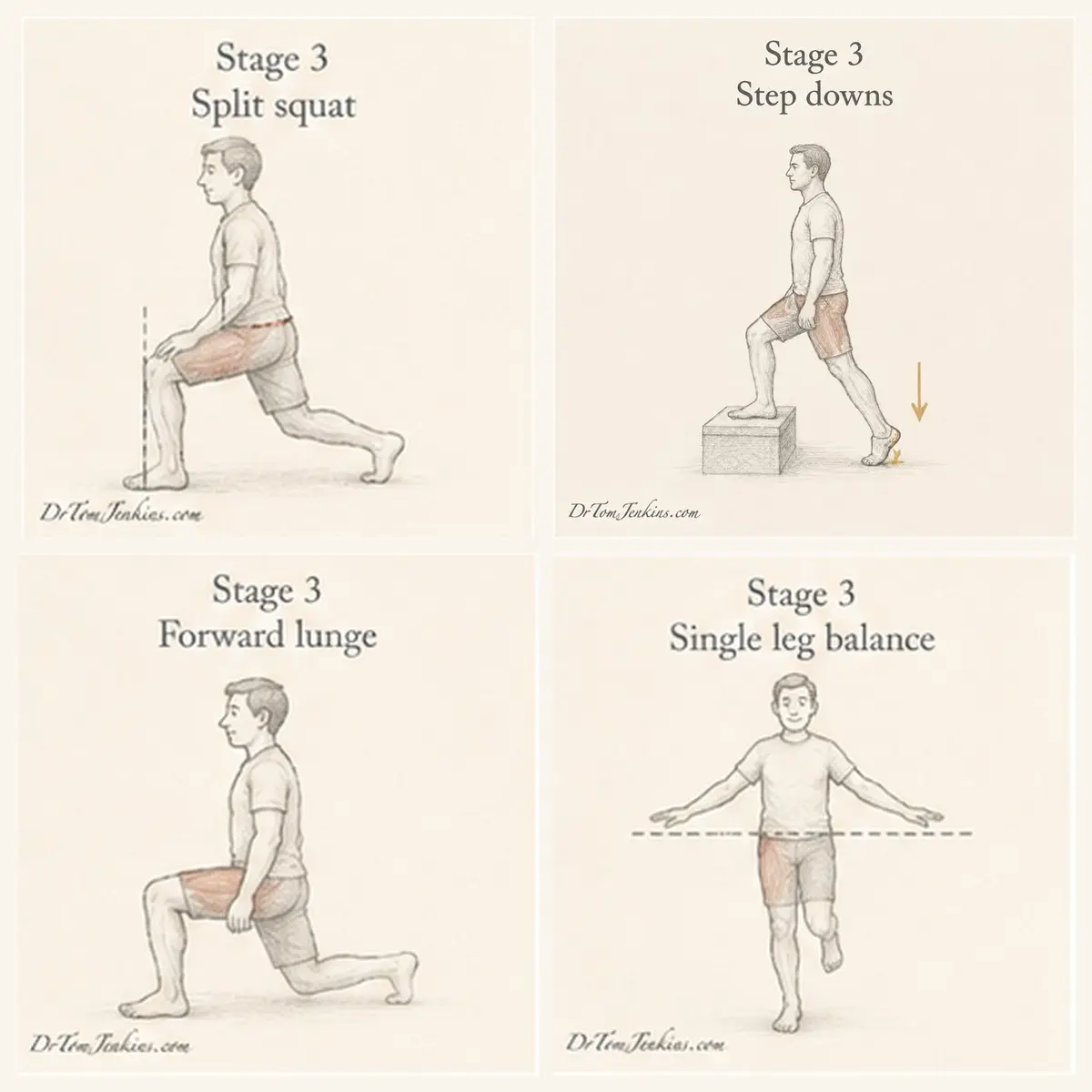
Split squat. One foot forward, one back, lower straight down, keeping the pelvis level and the knee tracking over the foot. Step downs. Stand on a step and lower slowly, taking the weight through the heel of the standing leg; this is demanding, so introduce it late. Forward lunge. Step forward into a lunge, torso upright, front knee tracking over the foot, then push back to standing. Single leg balance. Stand on one leg with the other foot just clear of the floor, keeping your pelvis level; progress by closing your eyes or standing on a softer surface. Once these are comfortable, return gradually to running or sport, increasing by no more than about 10 per cent a week.
Stretches
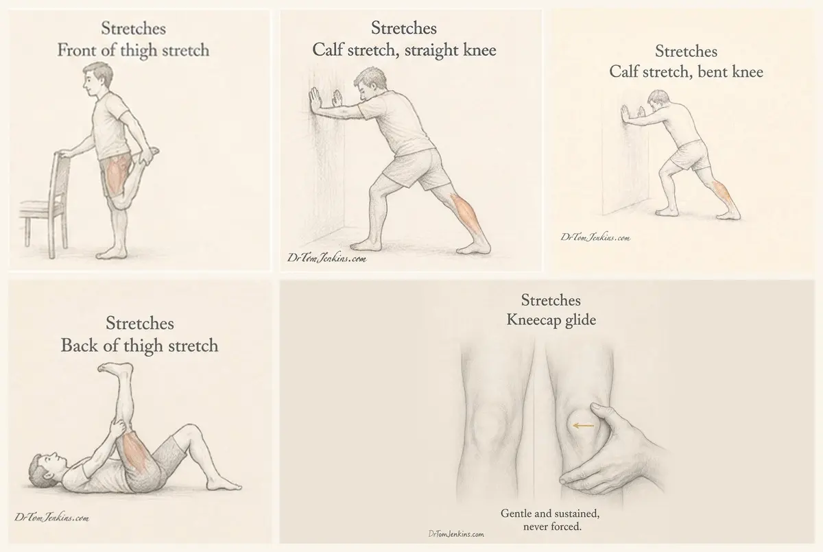
Hold each for 45 seconds, three times, once or twice a day. Front of thigh: standing, hold your foot behind you, tucking your bottom under; you should feel it in the thigh, not the knee. Calf, straight knee then calf, bent knee, both leaning against a wall with the heel down. Back of thigh: lying on your back, hold behind the thigh and straighten the knee towards the ceiling. Outer side of the kneecap: sitting with the leg straight and the thigh relaxed, use the heel of your hand to glide the kneecap gently towards the inner side and hold. Gentle and sustained, never forced.
Why it takes patience, and why the early exercises feel easy
Rebuilding strength and control is slow and repetitive, and this is where most people lose faith. I compare it to learning a musical instrument. A piano teacher tells you to practise regularly, and you take it on trust that the ability will come. You would not expect to play well after three weeks. The knee is the same: the results come from consistent repetition over months, on faith that change is happening before you can feel it. That willingness to keep going is the single biggest predictor of a good outcome.
The comparison is closer than it first sounds, because part of what you are doing is learning a skill rather than simply building a muscle. When you practise the piano you are not making your fingers stronger. You are teaching your nervous system a pattern until it runs without your attention. Rehabilitation has a good deal of that in it.
Which is why the early exercises can feel disappointingly easy. At that stage the aim is accurate repetition rather than exhaustion: the same movement, done well, often enough that it stops needing concentration. It is worth doing carefully, because a movement practised sloppily is still being learned.
Roughly, the sequence is: learn the movement, then strengthen the movement, then make it resilient. Early on, quality matters more than effort. In the middle the loads go up and the strengthening does the real work, and since that is the part with by far the strongest evidence behind it, it is not a stage to skip or rush. Later the movement should become automatic again, quicker, and more like the thing you actually want to do, whether that is a hill walk, a squash court or four flights of stairs at work.
One thing I want to be clear about. The goal is not to spend the rest of your life consciously managing your knee. It is precisely the opposite. The point of all the practice is to get back to not thinking about it at all.
What I usually do not recommend
Routine scans, for the reasons above. Steroid injections, which are not a treatment for straightforward patellofemoral pain; the British Patellofemoral Society takes the same position. Knee braces and straps for patellofemoral pain, where the evidence does not support them. Passive treatments used alone, such as ultrasound machines or electrotherapy. Surgery, which is rarely indicated and is reserved for a small minority with correctable structural problems.
When to seek help
See a clinician if the kneecap pain is severe or not settling, if it is stopping you doing what matters to you, or if you want a clear diagnosis and a plan aimed at your knee rather than knees in general. Some features point away from simple patellofemoral pain and are worth checking promptly: a knee that truly locks or gives way, significant swelling, pain following a distinct injury, pain that wakes you at night unrelated to activity, or fever with a hot, swollen joint.
Frequently asked questions
Is this the same as arthritis?
No. It is an overload-related irritation of the joint. Some people also have wear within the joint and the two can coexist, but having patellofemoral pain does not mean you have arthritis or that you will develop it. And if you have been told you do have arthritis behind the kneecap, that is not the end of the story either, as set out above.
Do I need an X-ray or MRI?
Usually not to explain the pain, because a scan cannot do that. Imaging is used to look for co-existing conditions when the assessment raises the question.
Should I stop running or exercising?
No. Modify rather than stop. Reduce the aggravating loads for a while, stay generally active, and build strength.
Is the clicking or grinding a problem?
No. It occurs in most pain-free knees and is not a sign of harm.
How long until I am better?
Think in months, not weeks, and expect a slow start.
Am I damaging my knee if it hurts while I exercise?
Not within the limits above: discomfort that settles within an hour or so and is no worse the next morning is expected.
Should I use a knee brace?
Generally no for patellofemoral pain. Taping, properly tailored, is a better short-term option alongside exercise.
For clinicians: evidence notes and key references
This article is written for patients, and colleagues are welcome to use or share it.
A note for physiotherapy colleagues in particular. Please do share this with patients on your waiting list if it is useful. It is written to cover the ground you would otherwise spend much of a first appointment on: that the joint is overloaded rather than damaged, that timescales are months rather than weeks, that load management and progressive strengthening are the treatment, and that adherence over the long term is what determines the outcome. It is deliberately not a substitute for your assessment and says so repeatedly, and the staged programme is adapted from the British Patellofemoral Society’s own patient leaflet with credit given.
It presents anterior knee pain as an umbrella with patellofemoral pain as the dominant cause, and treats excessive lateral pressure and patellofemoral instability as points on a spectrum rather than separate diseases, since they share aggravating activities, examination findings and first-line management.
It also deliberately acknowledges that the relative contribution of patellar tracking, lateral soft tissue tightness and vasti balance remains contested, because current sources genuinely disagree. BMJ Best Practice retains a mechanical model with patellar maltracking, iliotibial band tightness and reduced patellar mobility as contributors, and recommends taping, bracing and lateral soft tissue work. The BJSM consensus statements and the 2019 JOSPT clinical practice guideline place the emphasis on load management and combined hip and knee exercise therapy, and advise against bracing and against selective vastus medialis training. The 2025 Dutch multidisciplinary guideline, which is GRADE-based and the most recent of these, puts exercise therapy first after clinical diagnosis and classes gait retraining, taping, bracing, dry needling and manual soft tissue techniques as uncertain. The position taken here is that the relative importance varies between individuals, which is the argument for assessment and subgrouping rather than protocol-driven care.
The section on motor learning is framed as skill acquisition and adherence rather than as a claim that movement retraining corrects pathology, which the evidence does not currently support. Progressive loading remains presented as the main event.
Key references
Crossley KM, Stefanik JJ, Selfe J, et al. 2016 Patellofemoral pain consensus statement, 4th International Patellofemoral Pain Research Retreat, Manchester. Parts 1 and 2. Br J Sports Med 2016;50(14):839-852.
Collins NJ, Barton CJ, van Middelkoop M, et al. 2018 Consensus statement on exercise therapy and physical interventions to treat patellofemoral pain, Gold Coast. Br J Sports Med 2018;52(18):1170-1178.
Willy RW, Hoglund LT, Barton CJ, et al. Patellofemoral pain: clinical practice guidelines. J Orthop Sports Phys Ther 2019;49(9):CPG1-CPG95.
Ophey MJ, Bosch K, Khalfallah FZ, et al. Dutch multidisciplinary guideline on anterior knee pain: patellofemoral pain and patellar tendinopathy. Knee Surg Sports Traumatol Arthrosc 2025;33(2):457-469.
Barton C, Balachandar V, Lack S, Morrissey D. Patellar taping for patellofemoral pain: a systematic review and meta-analysis. Br J Sports Med 2014;48:417-424.
National Institute for Health and Care Excellence. Osteoarthritis in over 16s: diagnosis and management. NICE guideline NG226, 2022.
BMJ Best Practice. Patellofemoral pain syndrome.
Further reading
- British Patellofemoral Society: Information for patients
- NHS Inform: Patellofemoral pain syndrome
- NHS: Knee pain
- Versus Arthritis: Knee pain
Dr Tom Jenkins BSc MBChB MSc MRCGP MFSEM
Musculoskeletal GP with Expertise in Sport & Exercise Medicine
Member of the Faculty of Sport & Exercise Medicine (UK)
Practising in Gloucestershire